2022.07.02.23
Files > Volume 7 > Vol 7 No 2 2022

Asma Mohammad 1 and Mohammad Khalil2,*
1 University of Mosaul. Iraq
2University of Mosul; Iraq
* Correspondence: [email protected]
Available from: http://dx.doi.org/10.21931/RB/2022.07.02.23
ABSTRACT
The study included isolating and diagnosing the fungi found in dust samples from homes and buildings such as basements and walls containing fungal growth in separate areas from the left side of the city of Mosul in northern Iraq, such as Al-Ghufran neighborhood, Al-Mazare’ neighborhood, and Al-Mohandesin neighborhood during October and November, and the relationship of these fungi to human diseases, including allergies and asthma. The isolation results showed many fungal genera, including Cladosporium, Penicillium, Aspergillus, Alternaria and Trichoderma. The research aims to study the fungi Alternaria and Trichoderma, where the percentage of the presence of Alternaria in the wall sample containing the previous fungal growth was 28.57%. Whereas the percentage of the presence of Trichoderma fungus in dust and gypsum falling on the surfaces of poorly ventilated rooms in the cellars was 42.86%, and the molecular diagnosis of fungal isolates was carried out, as it was confirmed that there is a match with the standard strains found in the gene bank. The Alt a1 and Exp genes responsible for asthma were also examined and detected in fungal isolates using PCR technology and polymerase chain reaction; the new genes in both isolates were recorded. On behalf of both the supervisor and the researcher with international numbers in the global gene bank.
Keywords: Household dust, allergies and asthma, indoor environments, Alternaria and Trichoderma, Mosul local fungi.
INTRODUCTION
Fungi are ubiquitous in the air, and their composition is essential to human health 1 and poses a health threat, especially to immunocompromised patients 2 . House dust is an environmental measure commonly used as an indicator of exposure to human for fungi 3; 4 . Alternaria is among the airborne genera responsible for allergic rhinitis or asthma 5. In addition, some species of Trichoderma, which are filamentous fungi responsible for infection-causing deaths in up to 53% of patients with immunodeficiency 6. Fungi contain many known allergens, including Alt a1, one of the main fungal allergens present in the ubiquitous species. Alt a1 is a highly present allergen in Alternaria sp and other fungi that can produce Alt a1 allergen present in 95-99% of homes 7 . Therefore, the response to allergies, whether nasal allergy or asthma, can be considered natural problems Associated with the inhalation of fungi in the air 8. Exposure to fungi occurs through inhalation of dust 9; exposure to airborne germs has been linked to upper and lower respiratory diseases 10. Indoor fungal exposure has been associated with increased risk and severity of asthma and allergies. Asthma and respiratory allergies are chronic inflammatory diseases that affect the airways and are characterized by bronchial hyperactivity and altered airway obstruction, which leads to recurrent episodes of coughing, shortness of breath, chest tightness, and wheezing, the severity of which can vary over time 11; 12. Early exposure to microbes or air pollutants and asthma is complex, and this depends on several factors, such as the nature of exposure to the pathogen, how it occurs and when the genetic susceptibility of the host 13. These factors indicate the importance of the surrounding environment in developing and exacerbating asthma 14. Some health studies have revealed the presence of some fungi in the respiratory tracts, including those of the genera Cladosporium, Eurotium, Penicillium and Aspergillus. Other genera such as Candida have been detected but in low proportions 13.
MATERIALS AND METHODS
Dust samples were collected indoors, where dust from the air-conditioning filter, dust accumulated on furniture, and dust samples were collected in ground rooms with poor ventilation and lighting. Finally, dust samples were collected from the damp walls containing former fungal growth. Fungi were isolated in dust samples using the dilution and dishes method to obtain dilutions of 10-1, 10-2,10-3, then 1 ml of each dilution was transferred to sterile Petri dishes, and 10 ml of PDA medium was added to each dish in three replicates. These dishes are then placed in nylon bags for each dilution and incubated at 28 °C for 7 days. The numbers of developing fungal colonies were counted, then the most common fungi were purified by transferring a small portion of the outer edges of the fungal colony to plates prepared with a PDA medium and preserving them for diagnostic purposes.
Preparation of Potato Dextrose Agar (PDA)
It was prepared by dissolving (39) grams of prepared nutritional medium (Lab.M.Limited, UK) in a liter of distilled water, then placing the medium in a Glass Flask and sterilizing it in an autoclave and adding the antibiotic gentamycin, then pouring the medium into Petri dishes and leaving the medium until it solidifies for use it later. 15
Preparation of Potata Sucrose Broth (PSB)
Potato tubers were taken in the amount of 200 g, peeled and cut into small cubes, boiled in 500 ml of distilled water for 20 minutes, then filtered with a gauze cloth and 20 g of sucrose dissolved in it, then completed the volume to 1 liter with distilled water Then pour the medium into Petri dishes and leave the medium to solidify for later use 15.
DNA isolation
Several steps were followed based on the instructions of the company's "Genomic DNA mini Kit of fungi" From Geneaid Company for DNA extraction.
DNA molecular size estimation
DNA molecular sizes were estimated by performing electrophoresis of samples on an agarose gel using a molecular ladder; 100 base pairs, then mixing the DNA with the loading dye and running it in a 2% agarose gel for min.
Amplification of a highly conserved ITS region in fungi using PCR technology
The polymerase chain reaction was performed using universal primers that amplify the ITS regions in genomic DNA, Table (1).

Table 1. Universal fungal primers
Then the reaction tubes were inserted into the German-origin thermal cycler to conduct the reaction using the particular program for the reaction, as shown in the following table (2).

Table 2. Polymerase chain reaction (PCR) program for ITS region amplification
DNA extraction from agarose gel
The packets resulting from the PCR reaction were extracted from the gel to be purified and sent for nucleotide sequencing testing, depending on the analysis kit supplied by (Geneaid) company.
Detection of the presence of ATL1 a and EXP genes in the Fungal isolates using PCR technique
Both ATL 1 a and EXP genes were detected in the fungal samples. The DNA template was 4 μl (100 nanograms), and each gene-specific primer 1 μl (10 picomols) were added to the contents of the Master mix, as shown in table (3).

Table 3. Alta1 a, EXP genes primer
Then the reaction tubes were inserted into the thermocycler to conduct the amplification reaction, using the particular program for the reaction, as shown in the following table (4).
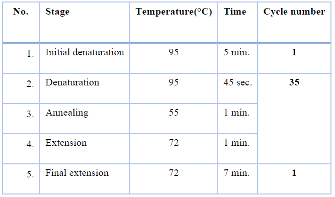
Table 4. Polymerase chain reaction (PCR) program for Alta1 a, EXP genes amplification
Determination Of Nucleotide Sequences For Amplified Pieces Using DNA Sequencing
The sequence of the nitrogenous bases of the fungi samples under study was determined. The PCR reaction products extracted from the gel were sent for the abovementioned samples. The sequence was read for the genes based on the 3130 Genetic Analyzer device supplied by the Japanese company Hitachi, and the sequences of the genes were matched with the sequences Genes documented in the National Center for Biotechnology Information NCBI the results were analyzed using BLAST software.
RESULTS
Isolation and diagnosis of fungi from dust samples
The results of isolating fungi from household dust samples showed the presence of different types of fungi (Cladosporium, Penicillium, Aspergillus, Alternaria and Trichoderma) and after purifying the most prevalent isolates and growing them on PD. The following fungal isolates were obtained in a medium (Fig 1 and adding table 5.

Table 5. Shows the percentage of races obtained and the regions from which they were isolated
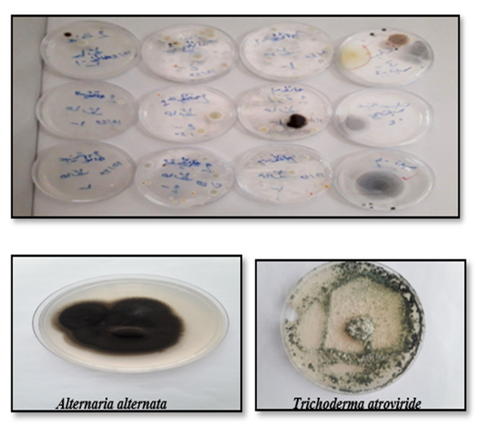
Figure 1. Fungi isolated from dust samples of some house's dust
Air pollution with fungal spores was of varying degrees in indoor environments, buildings and closed homes, as the largest number of fungi found in homes was recorded in dust samples collected from the surfaces of poorly ventilated ground rooms on the ground floor of some homes, and in gypsum samples falling from the walls of these rooms, followed by dust samples from walls containing previous fungal growth while, dust samples from air conditioner filters and dust accumulating on furniture recorded lower fungal growth, respectively. Fungal contamination of samples and surfaces differs from one house to another, and this mainly depends on the available conditions and factors, which vary according to the environment.
The results of isolation showed many fungal species present in the air of rooms and homes, Penicillium was the most common and frequent in a dust sample that was taken from a wall containing a previous fungal growth, where the percentage of its recurrence in the dishes was the highest and the percentage of its appearance in the dish was 71.43% Then followed by Alternaria, whose frequency was lower than Penicillium, and its appearance in dishes was 28.57%, and this was partially identical to what was isolated before 16; 17.
As for the dust samples of air conditioner filters, the frequency of Aspergillus fungus was the most prevalent, and the percentage of its appearance in dishes was 85.71%, while the percentage of other species that appeared in the dish was 14.29%, and this indicates that the air conditioner can cause the spread of fungal spores in house rooms. According to 18, fungal contamination of the air-conditioning filter, through which the air current passes, distributes the fungal spores throughout the room's atmosphere. Sometimes a foul smell or a cough occurs when the air conditioner is running, which is related to the fungal contamination inside the air conditioner as the air conditioners provide enough moisture to make the fungi grow and multiply on the filter.
As for the dust and gypsum samples falling on the surfaces of the ground rooms in the basements of some houses, several types were isolated, including Trichoderma, whose frequency was more common, and the percentage of its appearance in the dishes was 42.86%. In comparison, the appearance percentage of Aspergillus was 28.6%, the appearance of Penicillium fungi was 14.29%, and Cladosporium 14.31%. This indicates that the conditions of these rooms were a source of fungal contamination that made them rich in several similar fungal species. For those isolated by Lugauskas and Jaskeleviius 19 from the basement rooms of some buildings that were used as gymnasiums and some were used as book stores
DNA Extraction and PCR with Sequencing detection
Using a DNA extraction kit has several advantages, including the speed of extraction and obtaining high purity DNA. It also has a system capable of removing degrading enzymes as well as removing PCR inhibitors, thus resulting in high-quality DNA within a period not exceeding 60 minutes without resorting to the use of phenol or chloroform, and after the process of DNA isolation and extraction, electrophoresis was performed by adding of fungal DNA 1 μl in the presence of a DNA ladder in an agarose gel. The extracted fungal DNA samples were diagnosed as shown in the following figure )2). The results showed the amplification of ITS region for all samples and with high purity as shown in the following figure )2(.

Figure 2. Extraction of DNA from Genomic DNA, 1: Alternaria, 2: Trichoderma

Figure 3. Amplification of ITS region, 1: Alternaria, 2: Trichoderma
After the first fungus was isolated in pure form, PCR technology was performed to confirm its molecular diagnosis by investigating the duplication results representing the gene's DNA segment. The nitrogenous base sequences of the gene within the National Center for Biotechnology Information (NCBI) using the BLAST program, the name of the fungus for the isolate was obtained, and the sequences of the nitrogenous bases for the isolate gave the genus Alternaria alternata, which showed the percentage of match between the local isolate and the isolate recorded in the genebank by 100% as shown in the figure(4).
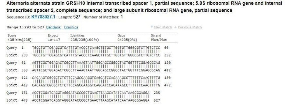
Figure 4. The sequences of nucleotides of Alternaria alternata
The result of the sequence of nitrogenous bases, the second isolate Trichoderma, was the Identities % between the local isolate and the isolate recorded in the gene bank was 99%, as shown in the following figure (5).

Figure 5. The sequences of nucleotides of Trichoderma atroviride
The results confirmed that techniques based on nucleic acid amplification could detect DNA quantities as they are usually fast and sensitive assays, thus overcoming the limitations of traditional diagnostics that are slow or have insufficient sensitivity, 20.
Detection of the presence of ATL 1 a and EXP genes in the fungi isolates
Specialized tools were used to detect and investigate the presence of ATL and EXP genes responsible for allergy and asthma in the selected local fungal isolates ( Alternaria and Trichoderma) by conducting multiplex polymerase chain reactions, The appearance of the bundles for each gene, where the length of the ATL 1 gene bundles is 510 base pairs, while the EXP gene bundles is 180 base pairs long. This confirms that the selected isolates contain their own gene that causes allergy and asthma, as shown in Fig (6) .

Figure 6 . Alt and EXP gene , 1,Altrenaria alternata, 2: Trichoderma atroviride
During the investigation and detection of ATL and EXP genes, new genes were globally registered in the International Information Bank (NCBI), where both isolates were registered with the name of both the supervisor and the researcher, the international number of the first isolate Alternaria LC632931 and the second isolate Trichoderma has the international number LC632932, and they were named MIK-AYJ1 and MIK-AYJ2 which is an abbreviation of the name of the supervisor and the researcher as shown in the fig (7) and fig (8).

Figure 7. Alternaria aletrnata NCBI BLAst

Figure 8. Trichoderma atroviride NCBI BLAst
Alternaria alternata has been significantly linked to asthma, and 21 have reported that Alternaria alternata is the most common allergen among those studied to date. It is considered one of the common biological pollutants in many countries and environments, it colonizes indoor environments, and the presence of these fungi in indoor environments makes people, especially those with immunodeficiency in these environments more susceptible to respiratory diseases, allergies and asthma, This is due to the ability of this fungus to produce Alt a1, which when inhaled provokes allergic reactions in patients allergic to mold. In this study, the Alt a1 gene was actually detected in Alternaria alternata, which was identical to researcher 3 . As for the inhalation of the spores of this fungus by people who suffer from asthma or allergies, this will lead to severe attacks of shortness of breath. In contrast, the Trichoderma plays an essential role in human health, as it is responsible for many diseases and causes death to reach to the highest levels of 53% in immunocompromised patients. Several potential human pathogens have been reported, such as Trichoderma atroviride, Trichoderma longibrachiatum, Trichoderma pseudokoningii and Trichoderma reese, which cause sinusitis, skin infections, pneumonia and stomatitis, as referring from 5 that reported the first confirmed case of lung infection. Due to the genus Trichoderma, which appears as invasive pulmonary aspergillosis in the patient, also, some people have been identified as having a respiratory allergy caused by Trichoderma fungus through skin prick tests of patients, where it was reported that the most critical respiratory allergen is Trichoderma harzianum 21.
CONCLUSIONS
There is a strong relationship between exposure to house dust fungi and asthma and allergies.
Household air conditioner filters contain a high percentage of respiratory fungi, including Cladosporium, Penicillium and Aspergillus
The extent of the importance of the polymerase chain reaction technology in detecting and investigating new genes and identifying mutations and strains for many organisms.
Fungi isolated from home contained Alta1 and Exp allergens.
The extent of the importance of air quality in the surrounding environment of humans.ACKNOWLEDGEMENT
The author is very thankful to the University of Mosul
for their facilities, which is helped the accomplishment of this work.
REFERENCES
1.Qi, Y., Li, Y., Xie, W., Lu, R., Mu, F., Bai, W., & Du, S. 2020). Temporalspatial variations of fungal composition in PM2. 5 and source tracking of airborne fungi in mountainous and urban regions. Science of the total environment, 708, 135027.
2. D’Amato, M., Molino, A., Calabrese, G., Cecchi, L., Annesi-Maesano, I., & D’Amato, G. 2018). The impact of cold on the respiratory tract and its consequences to respiratory health. Clinical and translational allergy, 8 1), 1- 8
3. Frankel, M., Timm, M., Hansen, E. W., & Madsen, A. M. 2012). Comparison of sampling methods for the assessment of indoor microbial exposure. Indoor air, 22 5), 405-414.
4. Leppänen, H. K., Täubel, M., Jayaprakash, B., Vepsäläinen, A., Pasanen, P., & Hyvärinen, A. 2018). Quantitative assessment of microbes from samples of indoor air and dust. Journal of exposure science & environmental epidemiology, 28 3), 231-241.
5.Nageen, Y., Asemoloye, M. D., Põlme, S., Wang, X., Xu, S., Ramteke, P. W., & Pecoraro, L. 2021). Analysis of culturable airborne fungi in outdoor environments in Tianjin, China. BMC microbiology, 21 1), 1-10.
6. Khalil, mohammad I. 2020."Identification of Cladosporium sp. Fungi by in- silico RFLP-PCR" Baghdad Sci.J, 17(1(Suppl.): 220-226.
7. Nastasi, N., Haines, S. R., Xu, L., da Silva, H., Divjan, A., Barnes, M. A., ... & Dannemiller, K. C. 2020). Morphology and quantification of fungal growth in residential dust and carpets. Building and Environment, 174, 106774. https://doi.org/10.1016/j.buildenv.2020.106774
8.Mashat, Bassam bin Hussein bin Hassan 2013). Does exposure to indoor fungi cause special diseases? Environmental and Health Research Department, Custodian of the Two Holy Mosques Institute for Hajj and Umrah Research, Umm Al-Qura University, Makkah Al-Mukarramah 1 16), 27-44.
9.Butte, W., & Heinzow, B. 2002). Pollutants in house dust as indicators of indoor contamination. Reviews of environmental contamination and toxicology, 175 1), 1.
10.Dannemiller, K. C., Gent, J. F., Leaderer, B. P., & Peccia, J. 2016). Indoor microbial communities: influence on asthma severity in atopic and nonatopic children. Journal of Allergy and Clinical Immunology, 138 1), 76-83.
11.Tipton, Laura, Elodie Ghedin, and Alison Morris. 2017. “The Lung Mycobiome in the Next-Generation Sequencing Era.” Virulence 8 3): 334–41.
12.Ichinose,M.;Sugiura,H.;Nagase,H.;Yamaguchi,M.;Inoue,H.;Sagara,H.;Tamaoki,J.;Tohda,Y.;Munakata,M.;Yamauchi,K. and Ohta,K. 2017): Japanese guidelines for adult asthma . Allergology International; 66:163– 189
13.Loss, G. J., Depner, M., Hose, A. J., Genuneit, J., Karvonen, A. M., Hyvärinen, A., ... & Ege, M. J. 2016). The early development of wheeze. Environmental determinants and genetic susceptibility at 17q21. American journal of respiratory and critical care medicine, 193 8), 889-897.
14.O'Driscoll, B. R., Hopkinson, L. C., & Denning, D. W. 2005). Mold sensitization is common amongst patients with severe asthma requiring multiple hospital admissions. BMC pulmonary medicine, 5 1), 1-10.
15.Alwan, Sabah Latif and Sukkar, Ahmed Rashid 2010). Test the ability of some fungal species in the biodegradation of some types of chemical pesticides. University of Kufa Journal of Life Sciences. 2 2):78-89.
16.Badran, B., Abid, H. A., & Ramadan, N. A. 2018). Isolation and Diagnosis Indoor/Outdoor Fungi of Schools in Tikrit City.
17. Sautour, M., Chrétien, M. L., Valot, S., Lafon, I., Basmaciyan, L., Legouge, C., … Caillot, D. 2018). First case of proven invasive pulmonary infection due to Trichoderma longibrachiatum in a neutropenic patient with acute leukemia. Journal de Mycologie Médicale. https://doi.org/10.1016/j.mycmed.2018.10.001
18.Hamada, N., & Fujita, T. 2002). Effect of air-conditioner on fungal contamination. Atmospheric Environment, 36 35), 5443– 5448. doi:10.1016/s1352-2310 02)00661-1
19.Lugauskas, A., & Jaskelevičius, B. 2007). Micromycetes Hazardous to Human Health in Buildings of Various Age and Use in Vilnius. Indoor and Built Environment, 16 4), 358–370. doi:10.1177/1420326x06081097 137.
20.Czurda, S., Smelik, S., Preuner-Stix, S., Nogueira, F., & Lion, T. 2016). Occurrence of fungal DNA contamination in PCR reagents: approaches to control and decontamination. Journal of clinical microbiology, 54 1), 148- 152.
21.Das, S., & Gupta-Bhattacharya, S. 2009). Trichoderma harzianum: occurrence in the air and clinical significance. Aerobiologia, 25 3), 137-145.
Received: 10 December 2021 / Accepted: 9 January 2022 / Published:15 May 2022
Citation: Asma Mohammad , Mohammad Khalil. Molecular identification of some allergenic fungi found in household dust in Mosul city. Revis Bionatura 2022;7(2) 23. http://dx.doi.org/10.21931/RB/2022.07.02.23